This atlas combines three presentations of cross-sectional anatomy - that of the dissecting room, CT and MRI. The series are matched to each other as closely as possible on opposite pages. Students of anatomy, surgeons, clinicians and radiologists should find the illustrations of anatomical cross-sections (obtained by the most modern techniques of preparation and photographic reproduction) and the equivalent cuts on imaging (obtained on state-of-the-art apparatus) both interesting and rewarding.

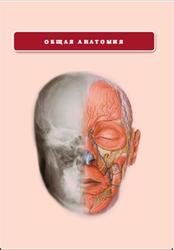
HEAD.
This section, at a deeper plane through the skull vault, demonstrates the falx cerebri (7), which is formed as a double fold of the inner, meningeal, layer of the dura mater (5) and which forms the dural septum between the cerebral hemispheres.
The inner layer of the dura is lined by the delicate arachnoid mater. The pia mater (9) is vascular and invests the brain, spinal cord, cranial nerves and spinal nerve roots. It remains in close contact with the surface of the brain, including the depths of the cerebral sulci and fissures.
Over the convexities of the brain, the pia and arachnoid are in close contact. Over the cerebral sulci and the cisterns of the brain base, the pia and arachnoid are separated by the subarachnoid space (8), which contains cerebrospinal fluid. This space is traversed by a fine spider's web of fibres (arachnoid: pertaining to the spider).
The total volume of cerebrospinal fluid in the adult is approximately 150 mL, of which some 25 mL is contained in the ventricular system, 25 mL in the spinal theca and the remaining 100 mL in the cerebral subarachnoid space.
CONTENTS.
Preface.
Introduction.
The importance of cross-sectional anatomy.
Orientation of sections and images.
Notes on the atlas.
References.
Acknowledgements.
Interpreting cross-sections: helpful hints for medical students.
BRAIN.
Series of Superficial Dissection [A–H].
Selected images.
3D Computed Tomograms [A–C].
HEAD.
Axial sections [1–19 Male].
Selected images.
Axial Magnetic Resonance Images [A–C].
Coronal sections [1–13 Female].
Sagittal section [1 Male].
TEMPORAL BONE/INNER EAR.
Coronal sections [1–2 Male].
Selected images.
Axial Computed Tomogram [A] Temporal Bone/Inner Ear.
NECK Axial sections [1–9 Female].
Sagittal section [1 Male].
THORAX.
Axial sections [1–10 Male].
Axial sections [1] Female Breast.
Selected images.
Reconstructed Computed Tomograms, Images [A–C].
Axial Computed Tomograms [A–D] Mediastinum.
Coronal Magnetic Resonance Images [A–C].
Reconstructed Computed Tomograms, Images [A–E].
Reconstructed 3D Computed Tomograms, Images [A–B].
ABDOMEN.
Axial sections [1–8 Male].
Axial sections [1–2 Female].
Selected images.
3D Computed Tomography Colonogram [A].
Coronal Computed Tomograms [A–C].
Axial Computed Tomograms [A–F] Lumbar Spine.
Coronal Magnetic Resonance Images [A–B] Lumbar Spine.
Sagittal Magnetic Resonance Images [A–D] Lumbar Spine.
PELVIS.
MALE – Axial sections [1–11].
Selected images.
Coronal Magnetic Resonance Images [A–C].
FEMALE – Axial sections [1–7].
Selected images.
Axial Magnetic Resonance Images [A–B].
Coronal Magnetic Resonance Images [A–C].
Sagittal Magnetic Resonance Image [A].
LOWER LIMB.
HIP – Coronal section [1 female].
Selected images.
3D Computed Tomograms Pelvis [A–B].
THIGH – Axial sections [1–3 Male].
KNEE – Axial sections [1–3 Male].
KNEE – Coronal section [1 Male].
KNEE – Sagittal sections [1–3 Female].
LEG – Axial sections [1–2 Male].
ANKLE – Axial sections [1–3 Male].
ANKLE – Coronal section [1 Female].
ANKLE/FOOT – Sagittal section [1 Male].
FOOT – Coronal section [1 Male].
UPPER LIMB.
SHOULDER – Axial section [1 Male].
SHOULDER – Coronal section [1 Male].
Selected images.
3D Computed Tomograms Shoulder [A–B].
ARM – Axial section [1 Male].
ELBOW – Axial sections [1–3 Male].
ELBOW – Coronal section [1 Female].
FOREARM – Axial sections [1–2 Male].
WRIST – Axial sections [1–3 Male].
WRIST/HAND – Coronal section [1 Female].
WRIST/HAND – Sagittal section [1 Female].
HAND – Axial sections [1–2 Male].
Index.
Бесплатно скачать электронную книгу в удобном формате, смотреть и читать:
Скачать книгу Атлас анатомии человека в срезах, Harold Ellis, Bari M Logan, Adrian Dixon, 2007 - fileskachat.com, быстрое и бесплатное скачивание.
Скачать pdf
Ниже можно купить эту книгу, если она есть в продаже, и похожие книги по лучшей цене со скидкой с доставкой по всей России.Купить книги
Скачать - pdf - Яндекс.Диск.
Дата публикации:
Теги: учебник по анатомии :: анатомия :: Harold Ellis :: Bari M Logan :: Adrian Dixon
Смотрите также учебники, книги и учебные материалы:

Следующие учебники и книги:
Предыдущие статьи: